- Medial Rectus Function
- Medial Rectus And Inferior Oblique Dmg
- Medial Rectus And Inferior Oblique Dmg Pain
Case
A 47-year-old patient presented to the hospital following an alleged assault involving multiple kicks to his face. He was found to have considerable ecchymosis around the right orbit, a right eye subconjunctival haemorrhage, and paraesthesia of the right side of his face. Visual acuity was better than 0.0 LogMAR in both eyes.
At his initial assessment he reported mild horizontal and vertical diplopia in upgaze and downgaze only. His examination demonstrated generalised mild restriction in all positions of gaze in the right eye, maximal in upgaze. In primary position only an exophoria was present. This corresponded with the Hess chart findings which consisted of a compressed pattern in the right eye and an expanded pattern in the left eye (Figure 1). On the basis of these findings there was concern about muscle or orbital fat entrapment. A CT performed at this time demonstrated a right orbital floor fracture with orbital fat herniating into the maxillary sinus (Figure 2). An orbital floor reconstruction was performed 10 days later without complication. After the orbital plate was inserted a forced duction test confirmed the globe was freely mobile.
Medial Rectus Function
The blended sheaths of the inferior rectus and oblique muscles then blend with the medial and lateral check ligaments, which are triangular sheet expansions of the medial and lateral recti muscles. Together, they form the suspensory ligament of the eyeball, a hammock-like sling that provides support to the eyeball. To investigate refractive changes after strabismus correction by combined recession of the Medial Rectus (MR) and Inferior Oblique (IO) muscles. We reviewed cases of combined MR and IO recession. Individuals with both preoperative refraction measurement and one month postoperative measurements were included.
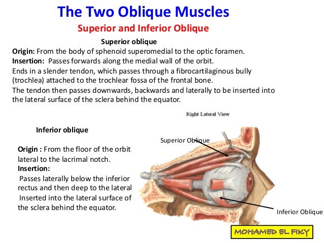
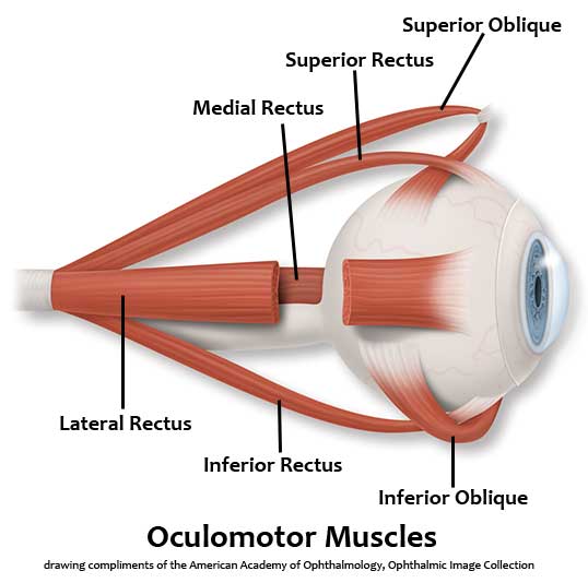
Typical space from limbus is: Medial rectus, 5 millimeters Inferior rectus, 6 millimeters Lateral rectus, 7 millimeters Superior rectus, 8 millimeter. Oblique Muscles. The oblique muscles of the orbit are superior and inferior. Their origin and insertion are as follows: Superior Oblique. Origin: from body of sphenoid superomedial to the optic. The other five extraocular muscles are the lateral rectus, superior oblique, superior rectus, inferior rectus, and the inferior oblique. Specifically, the medial rectus muscle works to keep the. Medial rectus - the ocular muscle whose contraction turns the eyeball medially medial rectus muscle, rectus medialis eye muscle, ocular muscle - one. Medial rectus - definition of medial rectus by The Free Dictionary.
Figure 1Preoperative clinical findings following initial injury. 1a. Ocular motility, 1b. Hess chart shows compression in the right eye.
Figure 2
CT scan of orbit using soft tissue windows. 2a Orbital floor fracture following initial injury. 2b. Coronal section following orbital floor repair. The bodies of the left and right medial recti have been highlighted to demonstrate their asymmetric positioning. The right medial rectus is clearly elevated in comparison to the contralateral side. 2c. Coronal section of the orbit shows the medial rectus tendon in close proximity to and distorted by the orbital plate. Anatomical structures are labelled, * indicates the globe. 2d. Sagittal section shows posterior extension of orbital plate. 2e. Coronal section following orbital plate exchange shows right medial rectus now symmetrical with left. 2f. Sagittal section following orbital plate exchange shows less posterior extension of orbital plate.
Immediately after surgery the patient complained of significantly increased vertical diplopia with new symptoms of torsion in all positions of gaze. He was found to have a right hypotropia and excyclotorsion maximal on levoversion of up to 10 degrees on synoptophore (Figure 3). There was also some limitation in upgaze that appeared to be due to mechanical restriction, particularly in dextroelevation.
Figure 3Postoperative clinical findings following orbital floor repair. 3a. Ocular motility, 3b. Synoptophore performed postoperatively with left eye fixating. Esotropia, excyclotorsion and mild left hypertropia noted in primary position. Left hypertropia maximal in dextroelevation. Esotropia was present in all positions of gaze and maximal in dextrodepression. Excyclotorsion was maximal in levoversion. 3c. Hess chart showing a compressed appearance in the right eye. Note restriction of upgaze in the right eye with associated overaction of the left superior rectus and underaction of right inferior oblique, medial rectus and inferior oblique.
A CT was performed, which showed changes in the position of medial rectus and inferior rectus following orbital plate insertion (Figure 2). The orbital plate elevated the right medial rectus and distorted the natural course of the muscle. When compared to the contralateral medial rectus, it was clear that the right medial rectus was elevated. Figure 2c demonstrates the medial rectus tendon appearing to ride on the border of the orbital plate. The inferior rectus is also seen in close apposition to the orbital plate. Based on imaging alone, entrapment or scarring between the muscle and plate could not be excluded. The superior rectus, lateral rectus, superior oblique and inferior oblique showed no abnormality on CT imaging.
Based on these clinical findings, combined with the CT, a diagnosis of medial rectus pulley distortion and possible inferior rectus mechanical restriction was made. The patient was offered a revision of the orbital plate to attempt to resolve the double vision. He elected to go ahead and 6 weeks after the injury the original plate was replaced with a smaller plate that extended less posteriorly and medially. No additional scarring or entrapment was found during the operation. After the plate was inserted the globe was freely mobile on forced duction testing.
Following his second operation the patient reported a total resolution of his symptoms. Examination revealed return of normal ocular motility and no significant torsion (Figure 4). The repeated CT scan showed return of the medial rectus to a normal position (Figure 2). He was able to resume all of his premorbid daily activities without any orthoptic aids.
Figure 4
Postoperative clinical findings following orbital plate exchange. 4a. Ocular motility, 4b. Synoptophore, left eye fixation, no torsion in any position of gaze, 4c. Hess chart.
Click to see full answer.
Then, what does the superior oblique muscle of the eye do?
The superior oblique muscle, or obliquus oculi superior, is a fusiform muscle originating in the upper, medial side of the orbit (i.e. from beside the nose) which abducts, depresses and internally rotates the eye. It is the only extraocular muscle innervated by the trochlear nerve (the fourth cranial nerve).
Additionally, is the inferior oblique muscle vertical or horizontal? When the eye is adducted, the oblique muscles are the prime vertical movers. Elevation is due to the action of the inferior oblique muscle, while depression is due to the action of the superior oblique muscle. The oblique muscles are also primarily responsible for torsional movements.
how do you test for superior oblique?
Clinical SignificanceInstead, as mentioned above, the superior oblique is tested by having the patient look down and in. By canceling the action of the inferior rectus muscle via contraction of the medial rectus, one can isolate the action of the superior oblique.


Medial Rectus And Inferior Oblique Dmg
What are the muscles that move the eye?
Medial Rectus And Inferior Oblique Dmg Pain
Eye muscle anatomy. There are six extraocular muscles that move the globe (eyeball). These muscles are named the superior rectus, inferior rectus, lateral rectus, medial rectus, superior oblique, and inferior oblique.